CT Scan
When symptoms point to a problem that cannot be fully understood through a physical examination or standard imaging, a CT scan may provide a clearer picture. This advanced imaging technique allows healthcare professionals to view detailed cross sectional images of structures inside the body.
A CT scan, short for computed tomography, combines X rays with computer processing to create detailed images of the body's internal structures. Unlike a conventional X ray, which produces a single two dimensional image, a CT scan can provide multiple views that help clinicians examine an area in greater detail.
CT imaging may be used to investigate injuries, infections, abnormalities, and other medical concerns when more detailed information is needed.
What Is a CT Scan?
A CT scan uses an X ray tube that rotates around the body while detectors collect information from different angles. A computer then processes this information to create detailed cross sectional images.
These images can be viewed individually or reconstructed into different planes, allowing healthcare professionals to examine internal structures from multiple perspectives.
Depending on the area being examined, a CT scan can provide useful information about bones, organs, blood vessels, and other tissues.
Why Might a CT Scan Be Recommended?
A CT scan may be recommended when your healthcare provider needs more detailed information than a standard X ray can provide.
It may be used to evaluate:
- Head and facial injuries
- Sinus and nasal conditions
- Bone and joint abnormalities
- Internal injuries
- Certain infections
- Tumors or abnormal growths
- Blood vessel conditions
- Complications involving internal organs
The reason for the scan determines which part of the body is examined and whether contrast material is required.
CT Scan for Head and Facial Conditions
CT imaging can be particularly useful when detailed information about the head and facial structures is needed.
For example, a CT scan may help evaluate certain facial injuries, sinus problems, skull abnormalities, or other conditions involving the head and neck.
It can provide detailed images of bony structures and may also help assess surrounding tissues depending on the type of examination performed.
CT Scan for Sinus Problems
When sinus symptoms are persistent, recurrent, or difficult to explain, a CT scan may sometimes be recommended.
A sinus CT can provide detailed images of the sinus cavities and surrounding structures. It may help healthcare professionals assess inflammation, blockage, structural abnormalities, or other changes that could contribute to ongoing symptoms.
CT imaging is not necessary for every case of sinusitis. Your healthcare provider decides whether it is appropriate based on your symptoms and examination.
What Happens During a CT Scan?
Before the scan, you may be asked to remove jewelry, glasses, or other metal objects that could interfere with imaging.
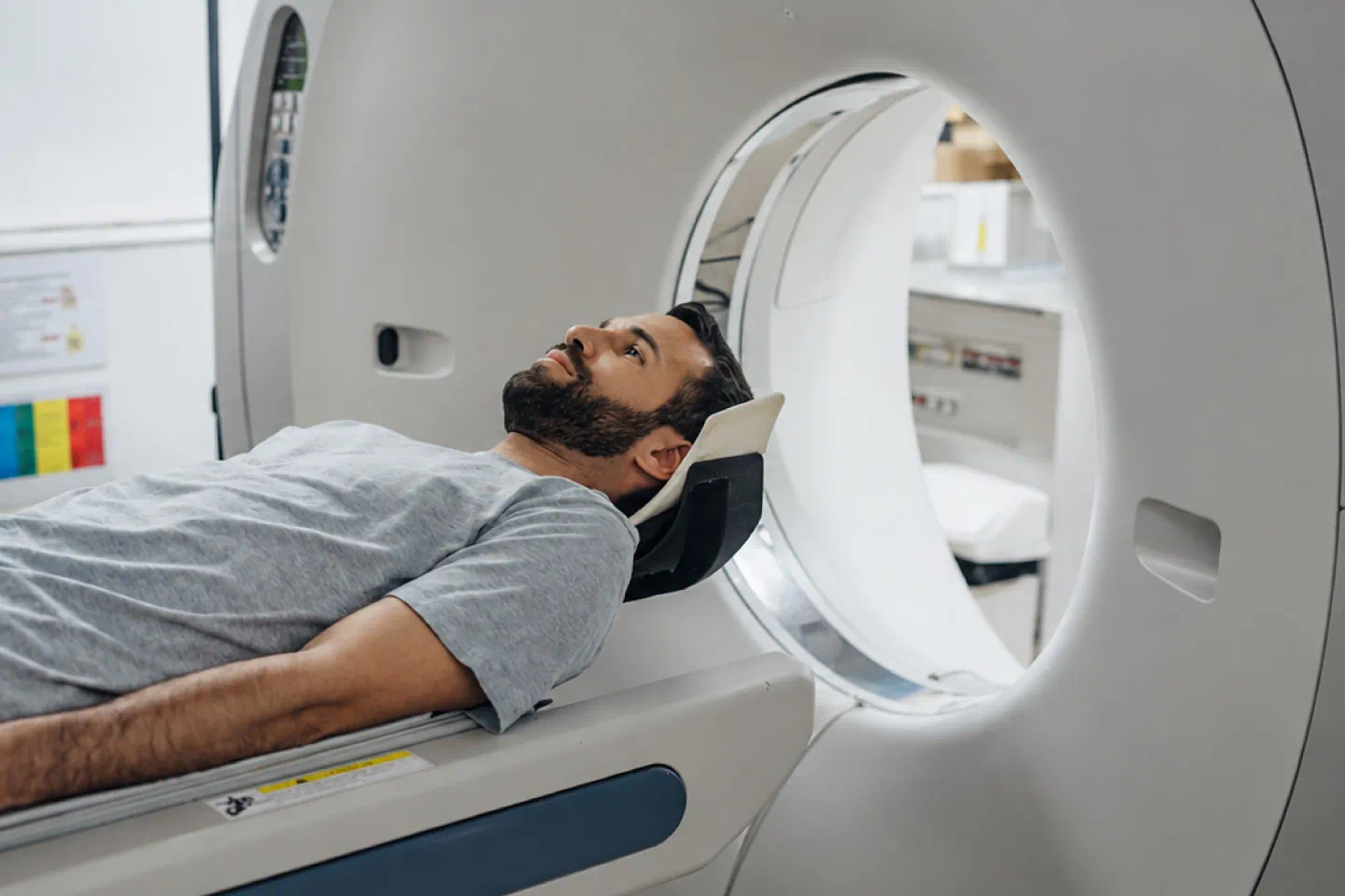
You will lie on a table that moves slowly through the opening of the CT scanner. The scanner itself is relatively open compared with some other imaging equipment.
You will need to remain still while the images are captured. Depending on the examination, you may also be asked to hold your breath briefly.
The scan itself is generally quick, although the total appointment time depends on the type of CT examination being performed.
Does a CT Scan Hurt?
A CT scan itself is painless.
You simply need to remain still while the scanner obtains the required images. If contrast material is needed, it may be administered through an injection or another method depending on the examination.
Some people experience a brief warm sensation or unusual taste after certain types of contrast are administered. Your healthcare team can explain what to expect beforehand.
What Is Contrast Material?
Some CT scans are performed without contrast, while others use contrast material to make particular tissues or blood vessels easier to see.
Contrast may be given orally or intravenously depending on what is being examined.
Before receiving contrast, tell your healthcare professional about any previous reactions to contrast material, kidney problems, or other relevant medical conditions. They can determine whether contrast is appropriate for you and whether additional precautions are needed.
How Is a CT Scan Different From an X Ray?
Both CT scans and conventional X rays use X rays to create images. The major difference is how the information is collected and presented.
A conventional X ray generally produces a two dimensional image of the examined area. A CT scan captures images from multiple angles and uses computer processing to create cross sectional views.
Because CT provides more detailed information, it may be recommended when a conventional X ray does not provide enough information for diagnosis.
Is Radiation Exposure a Concern?
CT scans use ionizing radiation, and the radiation exposure is generally higher than that of a conventional X ray.
Healthcare professionals consider the potential diagnostic benefit when recommending CT imaging and use appropriate protocols to keep radiation exposure as low as reasonably achievable while obtaining useful images.
If you are pregnant or think you may be pregnant, tell your healthcare provider before undergoing a CT scan. This allows the clinical team to consider the safest and most appropriate approach.
What Happens After a CT Scan?
After the images are obtained, they are reviewed by an appropriately trained healthcare professional, often a radiologist. The findings are then considered alongside your symptoms, medical history, and physical examination.
Depending on the results, your healthcare provider may recommend treatment, monitoring, additional imaging, laboratory tests, or referral to another specialist.
A CT scan provides important diagnostic information, but the images are interpreted in the context of your overall health.
When Might You Need a CT Scan?
A CT scan may be considered when your healthcare provider needs detailed information about a particular area of the body.
Whether you need one depends on your symptoms, medical history, examination findings, and the type of condition being investigated. Your healthcare provider can explain why CT imaging is being recommended and what they hope to learn from the examination.
Supporting More Accurate Diagnosis At Garv Diagnostic Centre & Hospital
A CT scan can provide detailed information when a conventional examination or X ray does not offer enough clarity. By creating cross sectional views of internal structures, CT imaging can help healthcare professionals investigate a wide range of conditions and make more informed decisions about further care.
If your healthcare provider recommends a CT scan, they can explain what the examination is intended to evaluate and whether any preparation is required.
Frequently Asked Questions
Q: How long does a CT scan take?
A: The actual scanning process is usually quick, but the total appointment time varies depending on the area being examined and whether contrast material is required.
Q: Is a CT scan painful?
A: No. The scan itself is painless. You simply need to remain still while the images are captured. If contrast is used, you may experience a brief sensation when it is administered.
Q: Can a CT scan detect tumors?
A: CT imaging can identify certain masses or abnormal areas and can provide useful information about their size and location. Additional testing may be needed to determine exactly what an abnormal finding represents.
Q: Is a CT scan the same as an X ray?
A: Both use X rays, but CT scans collect images from multiple angles and use computer processing to create detailed cross sectional views. This provides more information than a conventional X ray in many situations.
Q: Can I eat before a CT scan?
A: Preparation depends on the type of CT examination. Some scans require no special preparation, while others may require fasting, particularly when contrast material is being used. Follow the instructions provided by your healthcare team.
Q: Should I tell my doctor if I am pregnant before a CT scan?
A: Yes. Always inform your healthcare provider if you are pregnant or may be pregnant before undergoing CT imaging. Your provider can determine whether the examination is appropriate and consider suitable precautions.



Office Hours
Mon–Sat: Open 24 Hours
Sun: 8:00 AM–2:00 PM


©2026 | Garv Diagnostic Centre & Hospital | All Rights Reserved | Digital Marketing By Remedo